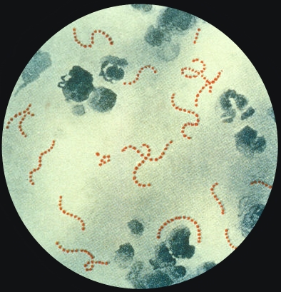
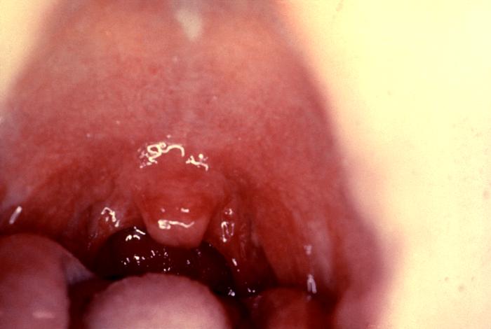
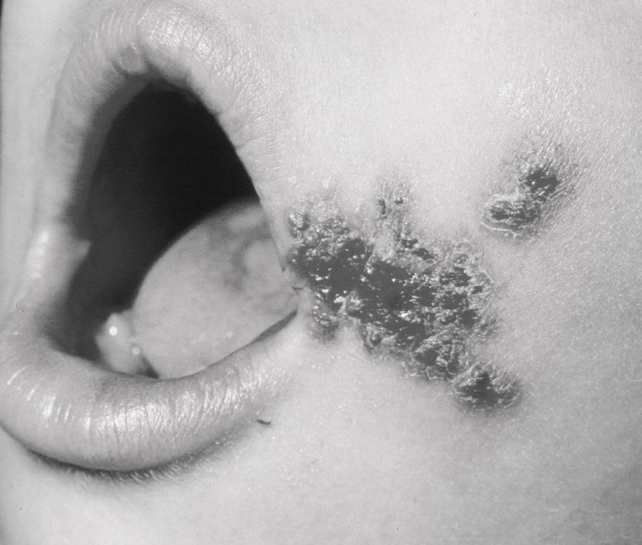
Streptococcus pyogenesOverview: Streptococcus pyogenes (also known as group-A streptococcus) is a Gram-negative, non-motile, non-spore-forming, catalase-negative, aerotolerant, opportunistic facultative anaerobe and a member of the Streptococcaceae family of bacteria (Figure 1). This organism has a capsule comprised of hyaluronic acid, protecting it from phagocytosis by neutrophils, and is beta-hemolytic. The key feature of beta-hemolytic species is that they produce an exotoxin called hemolysin, which has the ability to lyse red blood cells and subsequently generate clear zones of hemolysis on blood agar medium. Figure 1. Photomicrograph of Streptococcus pyogenes bacteria viewed using Pappenheim's stain. Last century, infections by S. pyogenes claimed many lives especially since the organism was the most important cause of puerperal fever and scarlet fever [900 X]. Virulence: S. pyogenes is an exogenous secondary invader that has various virulence factors, namely, M protein, which is displayed on the pathogen's surface and provides resistance to phagocytosis, lipoteichoic acid, which plays a role in attachment to target cells, the aforementioned carbohydrate-based bacterial capsule composed of hyaluronic acid, which surrounds the bacterium and allows the species to hide its own antigens and become unrecognizable by its host, the F protein, which also facilitates attachment to various host cells, numerous enzymes, including streptokinase, streptodornase, hyaluronidase, and finally exotoxins. M protein provides resistance to phagocytosis by inhibiting opsonization initiated by the alternative complement pathway. It does this by binding to host complement regulators. M protein found on some serotypes are also able to prevent opsonization by binding to fibrinogen. However, the M protein is also the weakest point in this pathogen's defense as antibodies produced by the immune system against M protein target the bacteria for engulfment by phagocytes. Moreover, the exotoxins generated by S. pyogenes are responsible for initiating fever, symptoms associated with the infamous strep throat, and scarlet fever rashes. There are various exotoxins produced, including streptolysin O, which is one of the bases of the organism's beta-hemolytic property, streptolysin S, which is a potent cell poison affecting many types of cell including neutrophils, platelets, and sub-cellular organelles, and streptococcal pyogenic exotoxin A (SpeA) and treptococcal pyogenic exotoxin C (SpeC), which are superantigens secreted by many strains of S. pyogenes and are responsible for the rash of scarlet fever and many of the symptoms of streptococcal toxic shock syndrome. Others virulence factors include streptokinase, an enzyme which enzymatically activates plasminogen, a proteolytic enzyme, into plasmin and, in turn, digests fibrin and other proteins, thus aiding in the invasion of wounds, and C5a peptidase, another enzmye which cleaves a potent neutrophil chemotaxin called C5a that is produced by the complement cascade of the innate immune system (Pier et al., 2004). C5a peptidase is necessary to minimize the influx of neutrophils early in infection as the bacteria are attempting to colonize the host's tissue. Finally, Streptococcal chemokine protease (ScpC), which is released by S. pyogenes and is responsible for preventing the migration of neutrophils to the spreading infection. ScpC degrades the chemokine interleukin-9 (IL-8), which would otherwise attract neutrophils to the site of infection. Pathogenicity: S. pyogenes is the cause of many human diseases, ranging from mild superficial skin infections to life-threatening systemic diseases. Infections typically begin in the throat or skin. Examples of mild S. pyogenes infections include pharyngitis (strep throat) (Figure 2) and localized skin infection (impetigo) (Figure 3). Erysipelas and cellulitis are characterized by multiplication and lateral spread of S. pyogenes in deep layers of the skin. S. pyogenes invasion and multiplication in the fascia can lead to necrotizing fasciitis (flesh-eating disease or flesh-eating bacteria syndrome), a rare potentially life-threatening condition requiring surgical treatment. Figure 2. Strep throat is caused by group-A streptococcus bacteria. These bacteria are spread through direct contact with mucus from the nose or throat of persons who are infected, or through contact with infected wounds or sores on the skin. Note the moderate redness of the oropharynx (soft palate) and tonsillitis caused by the pathogen. Figure 3. Impetigo is aa infectious sore cause by Streptococcus pyogenes. Infections due to certain strains of S. pyogenes can be associated with the release of bacterial toxins. Throat infections associated with release of certain toxins lead to scarlet fever. Scarlet fever rash first appears as tiny red bumps on the chest and abdomen, then spreads all over the body. It resembles a sunburn, and feels like a rough piece of sandpaper. It is usually redder in the axillary and groin areas. Similarly, puerperal fever can develop, a disease that is contracted by a woman during or shortly after childbirth, miscarriage or abortion, usually about the third or fifth day, that can develop into puerperal sepsis, which is a serious form of septicaemia. If untreated, it is life-threatening. Puerperal fever is diagnosed
when a woman shows a temperature above 100.4°
(38°C) over 24 hours or recurring from the end
of the first to the end of the tenth postpartum
day. An oral temperature of 100.4° F(38°C) or
more on any two of the first ten days postpartum
is also a warning sign. Some patients may report
a headache, vomiting, trouble breathing,
diarrhea, sore throat, or unusual vaginal
discharge as well. If caught early, puerperal
fever can be treated with antibiotics. When it
develops into puerperal sepsis, however, the
condition can lead to toxic shock syndrome,
multi-organ failure, and death. References: Pier, G.B., Lyczak, J.B., & Wetzler, L.M. (2004). Immunology, Infection, and Immunity. Washington: ASM Press. |
Beta-hemolytic: Beta hemolysis (β-hemolysis), sometimes called complete hemolysis, is a complete lysis of red cells in the media around and under the colonies: the area appears lightened and transparent. Compare with alpha-hemolysis.
|